
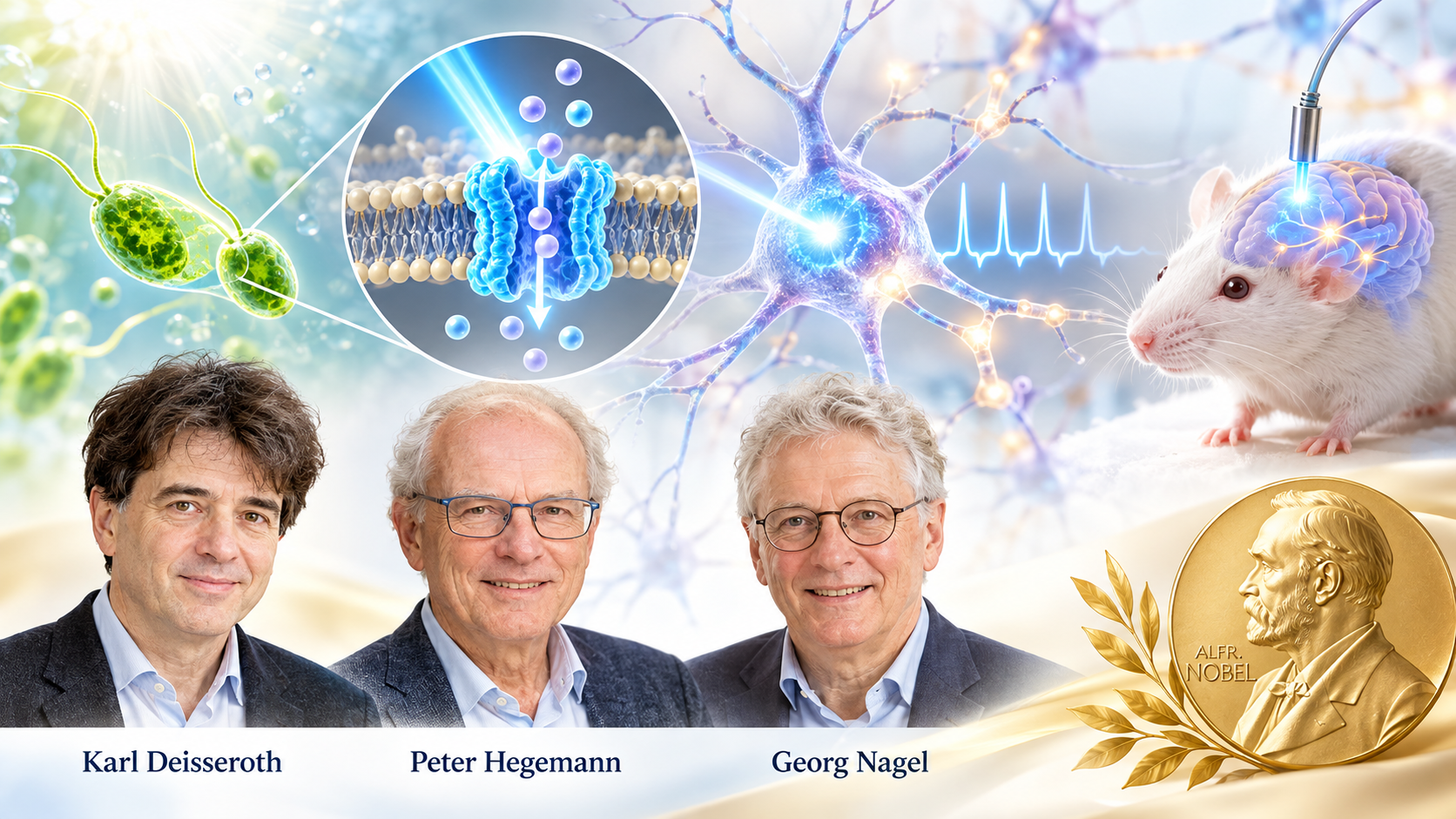
The 2026 Nobel Prize in Medicine and the Scientific Revolution That Began with a Microscopic Alga
How three scientists transformed a curious biological phenomenon into a technology that allows researchers to switch neurons on and off with light—and opened a new frontier in understanding memory, emotion, behavior, and the human mind.
Imagine a scientist sitting in a laboratory, watching a mouse move through a small enclosure. Nothing unusual happens until a tiny pulse of blue light reaches a particular group of neurons deep inside its brain. Suddenly, the animal stops moving. Its body becomes rigid, displaying a characteristic defensive response.
The light has not frightened the mouse in the ordinary sense. It has activated a neural circuit associated with fear.
Switch off the light, and the artificial stimulation ends.
What once belonged to science fiction has become a powerful experimental reality. Scientists can now select particular populations of brain cells, activate or inhibit them with extraordinary precision, and observe how those interventions change an animal’s behavior.
The technology is called optogenetics, and its development has earned Karl Deisseroth, Peter Hegemann, and Georg Nagel the 2026 Nobel Prize in Physiology or Medicine, awarded for their discoveries concerning light-gated ion channels and optogenetics.
The significance of this achievement extends far beyond neuroscience. It represents a fundamental change in how scientists investigate the relationship between the physical brain and the experiences it produces. For the first time, researchers acquired a practical method for controlling genetically selected neurons with pulses of light lasting only milliseconds.
Yet the story did not begin with a human brain, a neurological disease, or even a mammal.
It began with a microscopic green alga swimming toward sunlight.
The Tiny Organism That Changed Neuroscience
Deep within the biological world exists a single-celled organism called Chlamydomonas reinhardtii. It lives in freshwater environments and possesses an unusual ability: it can detect light and adjust its swimming direction accordingly.
For a creature without eyes, a brain, or a nervous system, this is a remarkable accomplishment.
How does a single cell recognize light? More importantly, how does it convert that recognition into movement?
These questions fascinated the German biophysicist Peter Hegemann, whose research helped uncover the molecular machinery behind this behavior.
The answer involved specialized proteins embedded in the organism’s outer membrane. These proteins, called channelrhodopsins, perform two functions simultaneously: they detect light and regulate the movement of electrically charged particles across a cellular membrane.
That combination would eventually transform the study of the brain.
To appreciate why, we must understand a basic principle of neuroscience.
Although the brain is composed of living cells, its communication depends heavily on electricity. Neurons maintain electrical differences across their membranes by controlling the movement of charged particles known as ions.
Sodium, potassium, calcium, and chloride ions are among the essential participants in this electrical activity.
When particular channels in a neuron’s membrane open, ions move across it, altering the cell’s electrical voltage. If the change reaches a critical threshold, the neuron generates an electrical impulse called an action potential.
These impulses travel through neural networks, helping coordinate everything from muscle movement and sensory perception to memory, attention, and emotion.
Normally, ion channels respond to electrical changes, chemical signals, or mechanical forces.
But channelrhodopsins possess an extraordinary property: light itself can open the channel.
When a photon strikes the protein’s light-sensitive molecular component, the protein changes its shape. This opens a passage through which ions can flow.
In other words, nature had already invented a molecular device capable of converting light into electricity.
The discovery raised a question that would alter the course of neuroscience: What would happen if this light-sensitive protein could be installed inside a neuron?
Giving Neurons a New Sense
Georg Nagel, working with Hegemann and colleagues, played a central role in identifying and characterizing channelrhodopsins as directly light-gated ion channels.
One of these proteins, Channelrhodopsin-2, commonly abbreviated ChR2, proved especially valuable.
When exposed to blue light, ChR2 opens and permits positively charged ions to cross the cell membrane. In a neuron, this can produce depolarization—the electrical change that brings the cell closer to firing an impulse.
The principle was deceptively simple.
A pulse of light could open a molecular gate. The gate could change the electrical state of a cell. And that electrical change could trigger neuronal activity.
The next challenge was to turn this molecular curiosity into a reliable method for studying the nervous system.
In 2005, Karl Deisseroth and his collaborators demonstrated that mammalian neurons genetically equipped with Channelrhodopsin-2 could be activated by light with millisecond precision.
The landmark experiment, reported by Edward Boyden, Feng Zhang, Ernst Bamberg, Georg Nagel, and Karl Deisseroth, established a practical foundation for modern optogenetics.
The researchers had accomplished something fundamentally new. They had introduced a gene encoding a light-sensitive protein into neurons, enabling those cells to respond to an external optical signal.
The term optogenetics captures this combination: optics, the science of light, and genetics, the science of genes.
Genetics determines which cells acquire the light-sensitive machinery. Light determines when that machinery is activated.
Together, they create a means of controlling selected neural populations with remarkable temporal precision.
This was not simply another technique for observing the brain. It was a way of intervening in its operations.
From Watching the Brain to Questioning It
For much of neuroscience’s history, scientists faced a fundamental problem: observing brain activity does not necessarily reveal what causes a particular thought, emotion, or behavior.
Suppose researchers discover that a group of neurons becomes active whenever an animal experiences fear.
Does that activity produce fear? Does it merely accompany fear? Or does it reflect the animal’s response to something threatening?
Observation alone cannot settle these questions.
Traditional methods provided partial answers. Electrical stimulation could activate brain tissue, but it often affected multiple types of cells simultaneously. Studying patients with brain injuries revealed important relationships between damaged regions and lost functions, but such injuries rarely isolated a single neural pathway.
Functional brain imaging later allowed scientists to observe patterns of activity associated with mental processes. Yet an image showing that a region becomes active during memory retrieval does not establish that the region independently generates the memory.
Optogenetics offered a different experimental approach.
Instead of merely asking which neurons become active during an experience, scientists could activate selected neurons themselves and observe the consequences.
If stimulating a particular circuit produced a defensive response, that provided evidence of its causal involvement in the behavior.
If inhibiting the circuit reduced the response, the interpretation became stronger.
Researchers could now manipulate neural activity with a degree of selectivity that had previously been extraordinarily difficult to achieve.
The distinction is crucial: correlation tells us what happens together; controlled intervention helps reveal what contributes to causing what.
This shift—from observation toward causal experimentation—is among the most consequential achievements of modern neuroscience.
Illuminating Memory, Fear, and Desire
The implications soon extended into some of the most mysterious territories of human experience.
Consider memory.
A memory is not stored in the brain like a photograph inside a filing cabinet. It involves changes in interconnected populations of neurons, whose patterns of activity and synaptic connections contribute to encoding, storing, and retrieving information.
Scientists have long attempted to identify the particular populations of neurons participating in individual memories. Such populations are often called engrams, although a memory generally depends on distributed networks rather than a single isolated cluster.
Using optogenetic techniques, researchers have tagged neurons active during particular experiences and later reactivated some of those populations with light.
In influential experiments involving mice, stimulation of neurons associated with a previously learned context could evoke memory-related behavioral responses even when the animal was no longer in that environment.
Other experiments demonstrated that manipulating neural activity during learning could alter subsequent associations.
These findings did not mean scientists had discovered a universal button for retrieving any memory. Nor did they establish that a complete subjective recollection could be reconstructed merely by illuminating a few cells.
They did, however, provide unusually strong evidence that specific neuronal populations participate causally in memory processes.
Similar methods have transformed the study of reward and motivation.
By stimulating particular dopamine-related pathways, scientists can investigate how neural circuits influence reinforcement, learning, and motivated behavior.
Other experiments examine the networks involved in hunger, sleep, movement, aggression, and defensive responses.
Each investigation brings researchers closer to understanding how activity in networks of physical cells contributes to the coordinated behavior of a living organism.
Yet an important distinction must remain clear: demonstrating that a neural circuit influences a behavior is not the same as explaining the complete subjective experience associated with that behavior.
A mouse freezing after stimulation of a defensive circuit does not establish exactly what, if anything, the animal consciously feels.
The technology reveals mechanisms, but interpreting those mechanisms requires scientific caution.
The Brain as an Orchestra
A useful way to understand the importance of optogenetics is to imagine the brain as an enormous orchestra.
Billions of neurons participate in intricate patterns of communication. Some become active, others remain silent, and still others modify the activity of neighboring cells.
A thought, movement, or emotional response emerges from the interaction of many components rather than the independent performance of one neuron.
Traditional brain imaging resembles listening to the entire orchestra and trying to determine which instruments contribute to a particular passage.
Electrical stimulation is somewhat like striking several instruments simultaneously.
Optogenetics makes it possible to select a particular group of performers and ask them to play at a specified moment.
Researchers can then observe how the rest of the orchestra responds.
But the analogy also exposes a limitation.
A violin does not produce the entire symphony, and a single neural population rarely explains an entire psychological experience.
The brain is a dynamic network in which timing, connectivity, chemical signaling, and previous experience all matter.
Optogenetics gives scientists a powerful means of studying that network, not a complete explanation of it.
Can Light Become a Medical Treatment?
The therapeutic possibilities are considerable.
Parkinson’s disease involves disturbances in circuits responsible for coordinating movement. Epilepsy involves abnormal patterns of neuronal excitation and synchronization. Depression, addiction, and several other psychiatric conditions are associated with changes in complex neural networks.
If scientists can identify the circuits contributing to these conditions, could precise manipulation of those circuits eventually improve treatment?
That possibility has motivated extensive research.
Unlike conventional drugs, which may influence receptors throughout the brain and body, an optogenetic intervention can be designed to target a more restricted population of cells.
In principle, such precision could reduce unwanted effects.
But translating the technology from experimental animals to human patients presents formidable obstacles.
First, most human neurons do not naturally contain the microbial light-sensitive proteins used in optogenetics. Researchers must introduce the relevant genetic instructions into the desired cells, often using modified viral vectors.
Second, visible light does not penetrate deeply into brain tissue. Reaching structures far below the surface may require implanted optical devices or other specialized light-delivery methods.
Third, long-term safety must be established. Gene expression, immune responses, tissue heating, cellular targeting, and the consequences of repeatedly manipulating neural circuits all require careful evaluation.
There is also the challenge of understanding which cells should be controlled.
A disease such as depression cannot simply be reduced to one defective switch. Its biology involves interacting neural circuits, genetics, stress responses, bodily physiology, and environmental influences.
Precision of stimulation is useful only when the underlying biological target is understood.
One particularly promising area of clinical investigation is vision restoration.
Because the retina is naturally exposed to light, it provides a comparatively accessible target for optogenetic approaches. Experimental treatments have attempted to make surviving retinal cells light-sensitive in people with severe photoreceptor degeneration.
Early human research has demonstrated limited but meaningful possibilities for restoring certain visual functions, although these methods remain investigational.
Thus, optogenetics has already revolutionized the laboratory study of disease, while its future role in routine clinical treatment remains to be established.
A New Window into Consciousness
Beyond medicine lies an even more difficult question: How does electrical activity in the brain give rise to subjective experience?
How do physical neurons participate in producing the sensation of pain, the recollection of childhood, the feeling of love, or the awareness of being oneself?
Philosophers have debated the relationship between mind and matter for centuries. Neuroscience has added increasingly detailed descriptions of the brain’s machinery, but the relationship between neural processes and conscious experience remains incompletely understood.
Optogenetics contributes an important experimental capability to this investigation.
Researchers can now test whether particular neural pathways are necessary or sufficient for specific measurable components of perception, attention, and behavior.
Combined with electrical recording, calcium imaging, advanced microscopy, and computational models, the technique allows scientists to examine how activity propagates through networks and how those patterns relate to an organism’s responses.
Artificial intelligence may further accelerate this work by helping researchers analyze immense datasets, identify patterns in neural activity, and construct models of circuit dynamics.
But we must resist an appealing exaggeration.
Controlling neurons is not equivalent to controlling consciousness, and identifying the circuitry of a behavior does not automatically explain the subjective experience accompanying it.
The brain’s electrical activity is indispensable to ordinary human consciousness, but precisely how organized neural processes generate experience remains an open scientific and philosophical problem.
Optogenetics provides a powerful instrument for investigating that problem. It does not settle it.
The Unexpected Power of Curiosity-Driven Science
Perhaps the most enduring lesson of this Nobel Prize concerns the nature of scientific discovery itself.
When researchers began investigating how microscopic algae respond to light, there was little reason to expect that the work would eventually transform neuroscience.
The original questions concerned the biology of a simple organism.
How does it detect illumination? What molecules participate? How does a cell translate environmental information into movement?
These were questions of fundamental curiosity, not immediate medical application.
Yet their answers revealed a molecular mechanism that could be transferred into an entirely different biological setting.
The light-sensitive machinery of an alga became a tool for investigating mammalian neurons.
The investigation of a single cell helped create a technology for examining the organization of complex brains.
This pattern appears repeatedly in the history of science. Discoveries made while studying bacteria, fungi, viruses, or obscure organisms have later reshaped medicine and biotechnology.
Nature often contains solutions to engineering problems that human beings have not yet learned to formulate.
Channelrhodopsins provide a particularly striking example.
Evolution developed these proteins for microbial responses to light. Scientists recognized their unusual properties and adapted them for an entirely new purpose.
The result was not the invention of a molecular switch from nothing, but the creative redeployment of a mechanism that already existed in nature.
The Light That Changed Our Questions
The 2026 Nobel Prize recognizes more than the discovery of an unusual protein or the invention of an ingenious laboratory technique.
It marks a turning point in humanity’s ability to investigate the physical foundations of behavior.
For centuries, scientists could observe the outward expressions of fear, desire, memory, and movement. Later, they learned to record the electrical signals accompanying those phenomena.
Now they can intervene in selected circuits and examine the consequences with extraordinary precision.
The advance does not make the brain simple. If anything, it reveals how deeply complex its operations are.
Thoughts are not isolated electrical sparks. Memories are not miniature pictures stored in single cells. Emotions are not produced by one universal switch.
They arise through interactions among neural populations, bodily states, prior experiences, and environmental conditions.
Optogenetics makes those interactions more experimentally accessible, while reminding us how much remains unknown.
There is something intellectually moving about the journey from a microscopic organism swimming toward sunlight to a laboratory in which light can influence the firing of neurons involved in memory, movement, and emotion.
The same physical phenomenon that guides a tiny alga through water has helped scientists develop a new language for questioning the brain.
And perhaps that is the deepest significance of this Nobel Prize.
Humanity has not yet learned how the brain transforms matter into subjective experience. But it has learned how to ask some of its most difficult questions with unprecedented precision.
The journey began with an organism searching for light.
It has brought us closer to understanding the organ through which we perceive light—and contemplate the universe that produces it.
Selected scientific references
- The Nobel Prize. (2026). The Nobel Prize in Physiology or Medicine 2026. NobelPrize.org. https://www.nobelprize.org/prizes/medicine/2026/
- Nagel, G., et al. (2003). Channelrhodopsin-2, a directly light-gated cation-selective membrane channel. Proceedings of the National Academy of Sciences, 100(24), 13940–13945. https://doi.org/10.1073/pnas.1936192100
- Boyden, E. S., Zhang, F., Bamberg, E., Nagel, G., & Deisseroth, K. (2005). Millisecond-timescale, genetically targeted optical control of neural activity. Nature Neuroscience, 8, 1263–1268. https://doi.org/10.1038/nn1525
- Deisseroth, K. (2011). Optogenetics. Nature Methods, 8, 26–29. https://doi.org/10.1038/nmeth.f.324
- Liu, X., et al. (2012). Optogenetic stimulation of a hippocampal engram activates fear memory recall. Nature, 484, 381–385. https://doi.org/10.1038/nature11028
- Sahel, J.-A., et al. (2021). Partial recovery of visual function in a blind patient after optogenetic therapy. Nature Medicine, 27, 1223–1229. https://doi.org/10.1038/s41591-021-01351-4
Editorial note: The account of fear-circuit activation is an illustrative synthesis of optogenetic research, not a description of one specific experiment. Clinical applications and claims about consciousness have been distinguished from established laboratory findings.